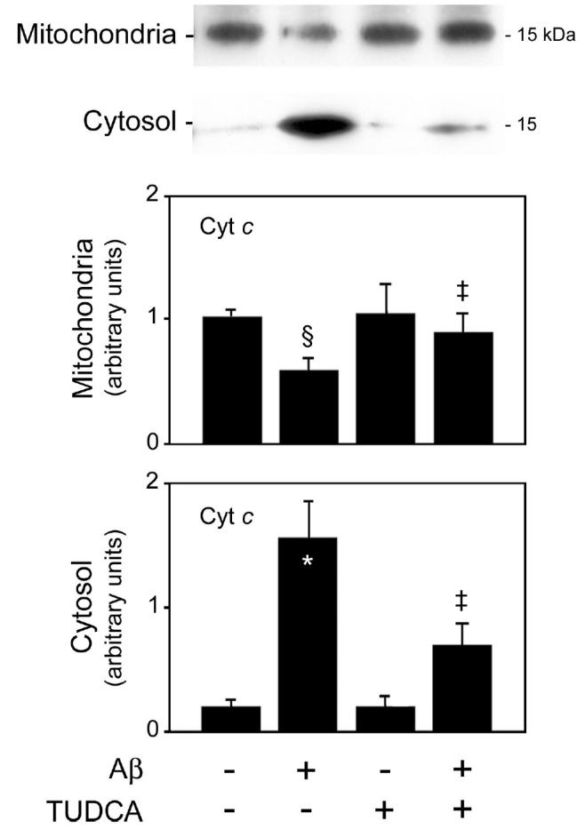
Figure 2.

TUDCA inhibits Aβ-induced cytochrome c release in rat cortical neurons. Cells were incubated with either vehicle (control), 25 μM Aβ fragment 25–35, 100 μM TUDCA, or a combination of Aβ plus TUDCA for 24 h as described in Materials and Methods. Mitochondrial (top) and cytosolic (bottom) proteins were processed for Western blot analysis. Following SDS-PAGE and transfer, the nitrocellulose membranes were incubated with a monoclonal antibody to cytochrome c (Cyt c). Histograms are mean ±SEM for at least 3 different experiments. §P < 0.05 and *P < 0.01 from control; ‡P < 0.05 from Aβ.
