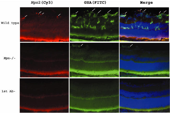
Figure 2.
Immunohistochemical staining for Npn2 is increased in hypoxic retina. Ocular sections from P17 wild type mice or npn2−/− mice with ischemic retinopathy were double–labeled for Npn2 (Cy3, red, 1st column) and the vascular cell marker, Griffonia simplicifolia lectin B4 (GSA; FITC, green, 2nd column). There was staining for Npn2 in tissue extending from the surface of the retina into the vitreous cavity (1st column, top row, arrows). Labeling with GSA (2nd column, top row) showed neovascularization on the surface of the retina (arrows) and retinal vessels within the retina. Merging of the 2 images (3rd column, top row) showed that the Npn2 expression was within the retinal neovascularization on the surface of the retina (arrows). Sections from npn2−/− mice with ischemic retinopathy rarely showed retinal neovascularization, but a section with a tuft of neovascularization that labeled with GSA was identified (2nd column, middle row, arrow). There was no staining for Npn2 within the neovascularization or elsewhere on the section (1st column, middle row). The bottom row shows a section adjacent to the one shown in the top row in which primary anti-Npn2 antibody (1st column) or GSA (2nd column) were omitted, and there is no staining.

