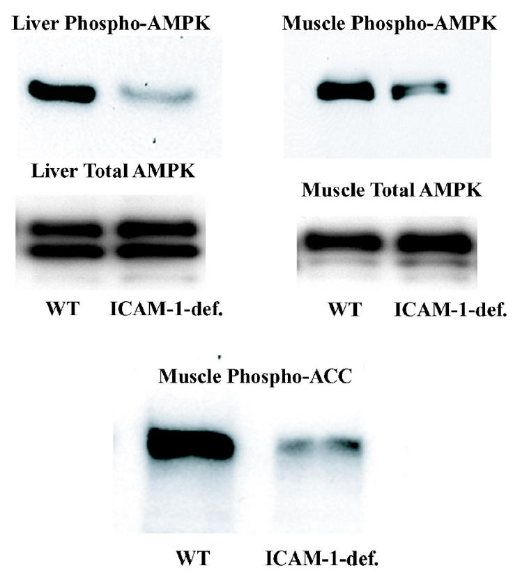
Figure 5.

Measurement of the phosphorylation status of AMPK and ACC in fasted WT and ICAM-1–deficient mice. Western blot analysis of total protein from soleus muscles and from livers of WT or ICAM-1–deficient mice fasted for 24 h. Protein extract was made from soleus muscles and livers of WT or ICAM-1–deficient mice sacrificed at the end of the dark cycle. Antibodies used were either anti-phosphoAMPK, anti-AMPK-α, or anti-phosphoACC as indicated. Each lane contains combined protein extract from 2 mice. Each experiment was repeated 2 times.
