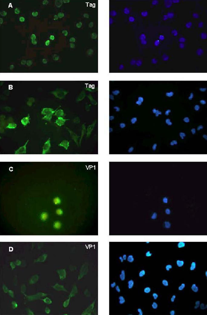
Figure 2.

Cellular localization of viral Tag and VP1 proteins in SV40-infected CV-1 cells (A, C) and in human SV40-immortalized fibroblasts MRC5-SV2 (B, D). Fixed cells were incubated with the Tag specific monoclonal antibody pAb101 (DBA Italia, Segrate, Italy) (A, B,) to localize the Tag, or with goat anti-SV40 serum (C, D) to localize the VP1. An adequate secondary antibody conjugated with fluorescein isothiocyanate (FITC) was used to reveal the signals (left column). Nuclei were visualized with 4′, 6-diamino-2-phenylindole (0.5 mg/mL) (Sigma, Milano, Italy) (right column). Left and right panels represent the same field analyzed for the 2 different dyes.
