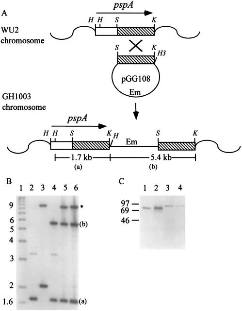
FIG. 3.
Construction and characterization of derivatives expressing altered PspA serotypes. (A) Construction of WU2 derivatives carrying an erythromycin resistance insertion downstream of pspA. WU2 was transformed with pGG108, which contains the 3′ 1.2 kb (▧) of WU2 pspA. Recombination resulted in insertion of the plasmid immediately downstream of pspA, with duplication of the 1.2-kb fragment (GH1003 chromosome). Chromosomal DNA from this strain was used to transform WU2 pspA into D39, with selection for erythromycin resistance following recombination in homologous sequences flanking pspA. The positions of HindIII (H), SacI (S), and KpnI (K) restriction sites are indicated. (a) and (b) indicate restriction fragments shown in the Southern blot in panel B. (B) Southern blot analysis. HindIII-KpnI-digested chromosomal DNA was hybridized with a full-length pspA-specific probe, as described in Materials and Methods. The complete D39 pspA gene (lane 3) is present on a single 2.1-kb HindIII-KpnI fragment (43). The 8.9-kb band (indicated by an asterisk) in lanes 3, 5, and 6 results from hybridization of the probe with the closely related pspC gene, which is present in D39 but not WU2 (35). WU2 pspA (lane 2) contains an internal HindIII site, as shown in panel A. Not shown is the 350-bp HindIII fragment, which was detected in WU2 and all derivatives containing WU2 pspA. Lane 1, kilobase ladder; lane 2, WU2; lane 3, D39; lane 4, GH1003 (WU2 with insertion downstream of pspA); lane 5, MA139 (D39 with WU2 pspA); lane 6, MA158 (D39 with WU2 pspA). (C) Western immunoblot analysis of cell lysates from derivatives expressing an altered PspA serotype. Blots were reacted with the PspA-specific monoclonal antibody Xi126. The positions of molecular mass standards (in kilodaltons) are indicated on the left. Lane 1, D39; lane 2, MA1025 (WU2 derivative expressing D39 PspA); lane 3, WU2; lane 4, MA139 (D39 derivative expressing WU2 PspA).

