
Figure 1.
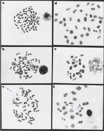
Metaphase spreads of male mice treated with ochratoxin A and aflatoxins showing: a) centromeric attenuation. b) deletion. C) chromatid break (small arrow), fragment (large arrow) of bone marrow cells and d) polyploidy. e) autosomal univalent. F) x-y univalent, of spermatocyte cells.