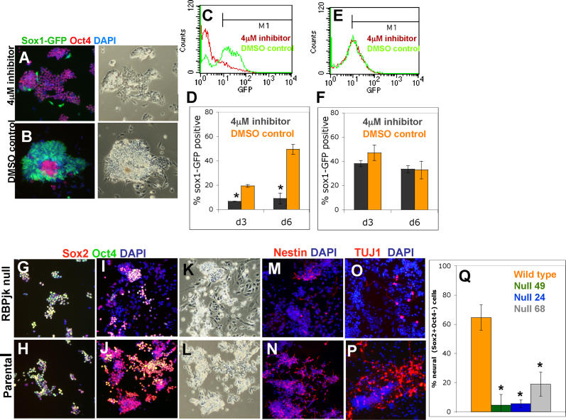
Figure 3. Notch Signalling Is Required for Efficient Neural Specification of ES Cells.
(A–F) Monolayer differentiation of 46C cells (A–D) or R26NotchIC cells (E and F) exposed to 4 μM γ-secretase inhibitor or to equivalent amounts of DMSO diluent. (A and B) Sox1GFP and immunostaining for Oct4 on day 5. (C and E) Typical FACS profiles for Sox1GFP on day 6. (D and F) Proportions of Sox1GFP+ cells (average of triplicates).
(G–P) Monolayer differentiation of RBPJk-null ES cells and the parental D3 ES cell line. Cultures were fixed and stained as indicated on day 2 (G and H) or day 6 (I–P).
(Q) Proportion of Sox2+ Oct4− cells on day 6 (average of duplicates).

