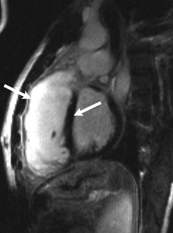
Figure 4.

Cardiac magnetic resonance with delayed contrast enhancement in a patient after tetralogy of Fallot repair. The areas with fibrosis have an increased signal intensity (white areas, left arrow) as compared with those of healthy myocardium (dark areas, right arrow)
