
Figure 1.
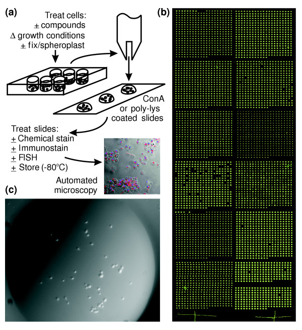
An overview of spotted cell microarrays. (a) Cell chips are constructed using slotted steel pins to print cells robotically from 96-well plates onto poly-L-lysine or Con A/Mn2+/Ca2+-coated glass slides. The sample image shows arrayed yeast cells immunostained for tubulin using fluorescein isothiocyanate (FITC)-conjugated-goat anti-rat IgG/rat anti-α-tubulin (red), overlaid on a bright field image and a DAPI-stained image (blue) of the cells' nuclei. FISH, fluorescence in situ hybridization. (b) Wide-field light scattering image of a cell microarray (approximately 2 cm × 6 cm) containing around 4,800 viable, haploid yeast deletion strains. The bright dots arise from light scattered when scanning the array with a Genepix DNA microarray scanner. Spots are around 200 μm in diameter, separated by 410 μm. (c) Close-up of a typical spot from the microarray showing distinct cells at 40× magnification. This image was taken immediately after printing, so growth medium (YPD, 17% glycerol, 200 mg/l G418) is still visible.