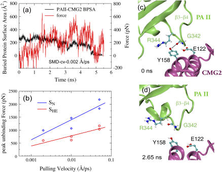
FIGURE 6.
Forced detachment of PAII from the CMG2 binding pocket in SMD simulations of the SHE structure. (a) Profiles of applied force and BPSA between PAII and CMG2 from a representative 0.002 Å/ps cv-SMD simulation. (b) Comparison of peak forces required to unbind PAII from CMG2 between the SN and the SHE structures in cv-SMD stretching simulations with various constant velocities. Significantly lower force is required to detach PAII when the Arg-122PA-Glu-122CMG2 salt bridge is broken under low pH conditions. (c–d) Snapshots of the PAII-CMG2 binding interface obtained from the representative SMD simulation.

