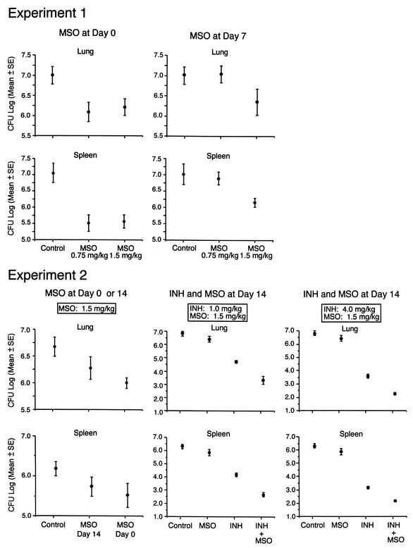
FIG. 3.
Growth of M. tuberculosis in the lungs and spleens of guinea pigs after M. tuberculosis challenge. At the end of the observation period, the animals described in the legend to Fig. 2 were euthanized, and the numbers of CFU of M. tuberculosis in the right lung and spleen were assayed. Samples from the few animals that died before the end of the observation period were cultured immediately after death. Data are the mean and standard error (SE) for all animals in a group (Table 3). The lower limit of detection was 2.0 log units per organ (1 CFU on a plate seeded with an undiluted 1% sample of an organ, i.e., 100 μl of a total sample volume of 10 ml). In experiment 2, two lung cultures and four spleen cultures from the group treated with INH (4.0 mg kg−1 day−1) plus MSO (1.5 mg kg−1 day−1) had 0 CFU on plates seeded with undiluted samples. For statistical purposes, these organs were scored as containing 2.0 log units.

