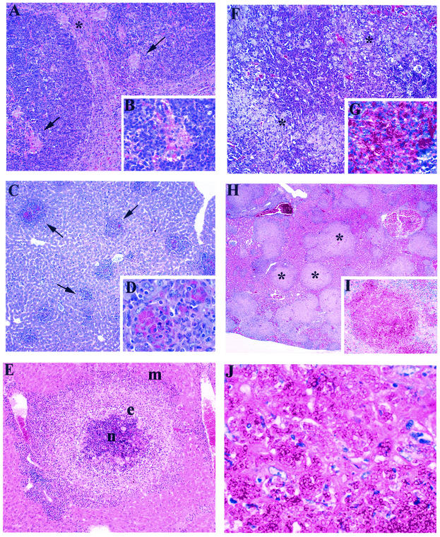
FIG. 5.
Composite illustration of the main histological findings in the livers and spleens of wild-type and GKO BALB/c mice infected with P. marneffei. Mice were injected i.v. with 3 × 10⁵ conidia and sacrificed at different times postinfection for histological analysis. (A) Wild-type BALB/c mouse spleen section at day 14 postinfection. Isolated single small granulomas were seen within the white pulp of the spleen. The asterisk indicates a granuloma at the edge; the arrows indicate granulomas within follicles. HE stain. Magnification, ×10. (B) P. marneffei yeast cells within macrophages. HE stain. Magnification, ×100. (C) Wild-type BALB/c mouse liver section at 7 days postinfection. Numerous small granulomas (arrows) are present. PAS stain. Magnification, ×4. (D) P. marneffei yeast cells within the macrophage cytoplasm. PAS stain. Magnification, ×100. (E) BALB/c mouse liver section showing a large granuloma with a target appearance; the central necrotic area (n) is circled by epithelioid histiocytes (e), macrophages (m), and granulation tissue. HE stain. Magnification, ×40. (F) GKO BALB/c mouse spleen section. The spleen structure is subverted by masses of macrophages loaded with yeast cells (asterisks). HE stain. Magnification, ×10. (G) Macrophages from GKO spleen cells filled with yeast cells. PAS stain. Magnification, ×100. (H) GKO BALB/c mouse liver section. Round masses of macrophages (asterisks) largely replace the liver parenchyma. HE stain. Magnification, ×4. (I) Macrophages from GKO liver cells filled with yeast cells. PAS stain. Magnification, ×100. (J) Macrophage cytoplasm occupied by a large number of yeast cells. PAS stain. Magnification, ×100.

