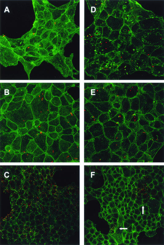
FIG. 4.
Confocal microscopy of TIB-73 cells infected (100 bacteria/cell) with EGD-e (A, B, and C) or lpeA mutant (D, E, and F). Cells were observed at time zero (A and D) and 2 h (B and E) and 4 h (C and F) postinfection. F-actin was stained with phalloidin (green). Bacteria were labeled with an anti-Listeria serum (red). At time zero, no difference between the two strains was observed. After 2 h, many wild-type bacteria polymerized actin and started forming comets (B), whereas the majority of mutant bacteria were not associated with actin (E). After 4 h, wild-type bacteria massively spread to the entire cell monolayers (C). In contrast, the cell monolayers remained poorly infected by the mutant, except for rare clusters shown in panel F (indicated by arrows), in which mutant bacteria polymerized actin and multiplied inside cells. Magnifications, ×480 (A, B, D, and E) and ×320 (C and F).

