FIG. 2.
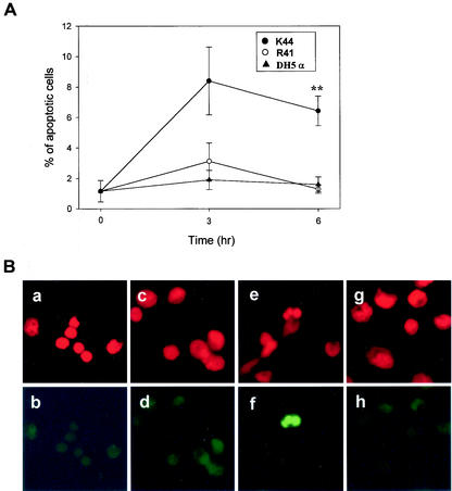
Clinical isolate K44 induces apoptosis in mouse peritoneal macrophages in vivo. Mice (n = 4 for each time point) were s.c. injected in the lower right flank with 107 bacteria suspended in 0.5 ml of PBS. Peritoneal macrophages were isolated at the indicated times after injection of bacteria. (A) Apoptotic cells were detected by the TUNEL method as described in the text. Data are presented as means ± standard errors (n = 4). **, P < 0.01 (K44 or R41 versus DH5α) according to Student's t test. (B) Nuclear morphology of mouse peritoneal macrophages stained with propidium iodide and the TUNEL method. Peritoneal macrophages were isolated at 6 h after the injection of bacteria. All of the cell nuclei in the field were stained with propidium iodide (a, c, e, and g). Apoptotic cells were stained by the TUNEL method (b, d, f, and h). Normal nuclear morphology was observed in control cells (a), in the cells of E. coli DH5α-injected mice (c), and in the cells of R41-injected mice (g). In these cases, cells were not stained by the TUNEL method (b, d, and h). In contrast, nuclear fragmentation with typical apoptotic morphology was observed in cells from mice injected with clinical isolate K44 (e and f).

