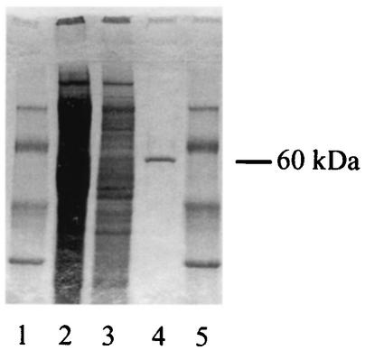
FIG. 3.
Purification of a UDP-glucosyltransferase from L. pneumophila. Samples were subjected to SDS-PAGE and stained with Coomassie brilliant blue R250. Lanes 1 and 5, molecular mass markers; (from top to bottom, 94, 67, 43, and 30 kDa; Amersham Biosciences); lanes 2 and 3, ultrasonic lysate of L. pneumophila Philadelphia I containing 30 and 15 μg of protein, respectively; lane 4, purified, 60-kDa UDP-glucosyltransferase (ca. 0.5 μg).

