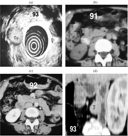
Figure 5.
(a) A well defined mass (Tn) of mid echogenicity seen lateral to the head of the pancreas on EUS in a patient with biochemical evidence of Zollinger–Ellison syndrome. (b, c) CT scans in 91 and 92 had failed to detect the tumour (T) but on repeat scanning (d) the lesion was clearly seen in the duodenal wall on a reconstructed sagittal view.

