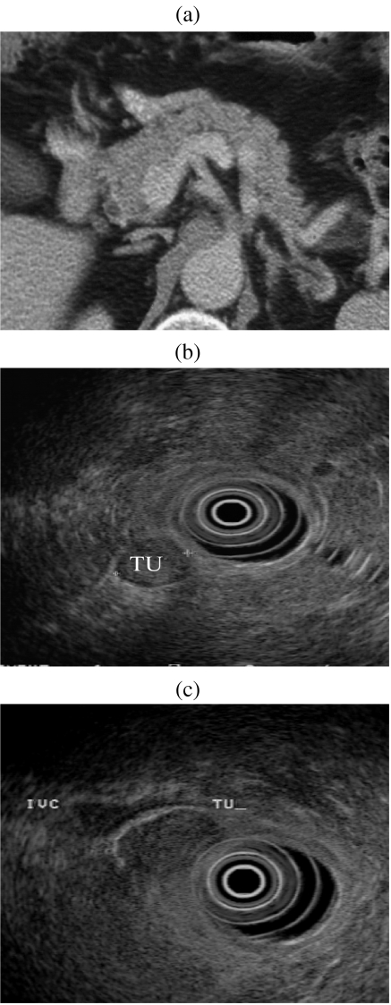
Figure 7.
(a) CT scan of pancreas reported as normal in a patient with biochemical evidence of a gastrinoma. (b, c) EUS demonstrates two small tumour nodules inferior to pancreatic head. (d, e) Review of the CT reveals two small enhancing nodules corresponding to the EUS appearance. Histology reveals two foci of malignant gastrinoma in peri-pancreatic nodes.

