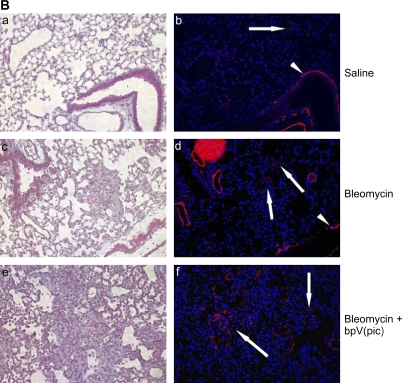
Figure 8.
Inhibition of PTEN worsens experimental fibrosis. (A) C57Bl/6 mice administered intratracheal bleomycin were treated in the presence or absence of the PTEN inhibitor bpV(pic). Animals were killed 21 d after bleomycin treatment, and lungs were assessed for total collagen. Mice treated with intratracheal saline were used as controls (n = 8 for each experimental group). The figure is representative of two separately performed experiments. (B) a and b: Lung sections from a mouse receiving intratracheal saline. c and d: Lung sections from a mouse receiving intratracheal bleomycin. e and f: Lung sections from a mouse receiving intratracheal bleomycin and intraperitoneal bpV(pic). Left panels (a, c, e): Trichrome stain. Right panels (b, d, f): Immunofluorescent stain for α-SMA (red) and nuclear DAPI stain (blue). Arrows are pointing to individual cells or clusters of myofibroblasts staining positively for α-SMA. Arrowheads identify α-SMA–expressing smooth muscle cells lining large airways. α-SMA–expressing myofibroblasts colocalize to fibrotic regions of the lung (200× original magnification).


