
Fig. 5.
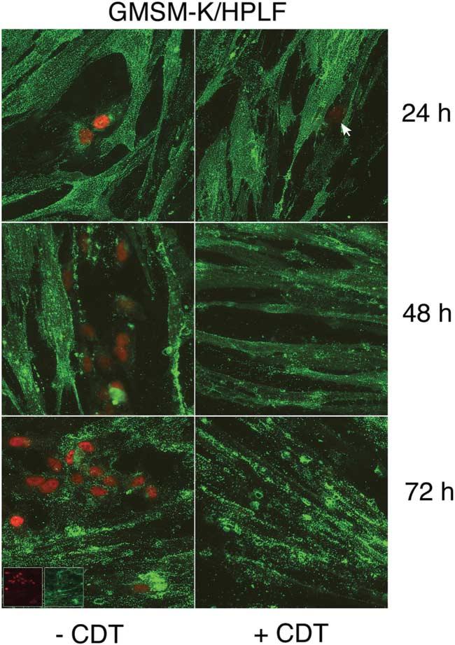
Time course of untreated and CDT-treated co-cultures of HPLFs and oral epithelial cells. HPLFs and GMSM-K cells were grown for 24 h on chamber slides and left untreated (left column) or treated with CDT (right column). Cultures were incubated and examined at 24, 48 and 72 h post-intoxication. Co-cultures were labelled with anti-SV40 T-antigen biotin-labelled mAb and streptavidin–Texas red-X (red fluorescence), and Ab-1 and Alexa Fluor 488 conjugate (green fluorescence). The arrow marks the position of a labelled GMSM-K cell in a CDT-treated co-culture. Immunofluorescence of the individual fluorochromes is shown in the insets in the lower left panel. Images are representative of multiple microscopic fields examined. Magnification ×60.