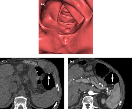
Figure 1.
Fecal tagging of stool with barium. (a) Endoluminal viewing shows two polypoid lesions. The true nature of these lesions can not be assessed with certainty. (b) Axial 2D source image of prone scan (the image is flipped to facilitate comparison with the scan in the supine position) shows fecal material to be very hyperdense (arrow), which allows easy differentiation from true polyps. (c) Axial image of supine scan shows mobility of the ‘polyp’ (arrow).

