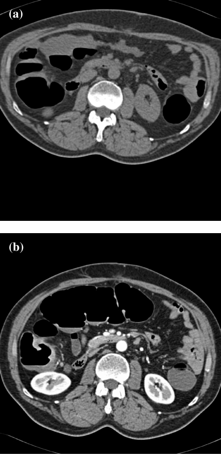
Figure 2.
Distension of the transverse colon: prone vs. supine imaging. (a) In the prone position, the transverse colon is collapsed and cannot be adequately assessed (the image is flipped to facilitate comparison with the scan in the supine position). The ascending and descending colon are adequately distended. (b) In the supine position, there is good distension of the transverse colon to assess the mucosa.

