Fig. 1.

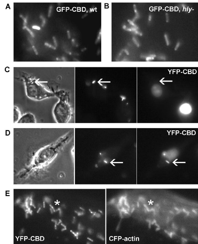
YFP-CBD as a marker of Lm delivery into cytosol.
A and B. Epifluorescence images of (A) wild-type Lm and (B) hly Lm decorated with HGFP-CBD after incubation with the purified fluorescent protein.
C and D. Phase-contrast (left), SNARF-labelled Lm (middle) and YFP-CBD fluorescence (right) images. (C) Cells viewed at 10 min after infection contained bacteria in phase-bright vacuoles (arrows) which were not labelled with YFP-CBD. (D) Cells viewed 60 min after infection showed some bacteria labelled with YFP-CBD (arrows).
E. YFP fluorescence image of a RAW 264.7 macrophage showing YFP-CBD-positive cytosolic Lm (left) with corresponding labelling by CFP-actin (asterisks).
