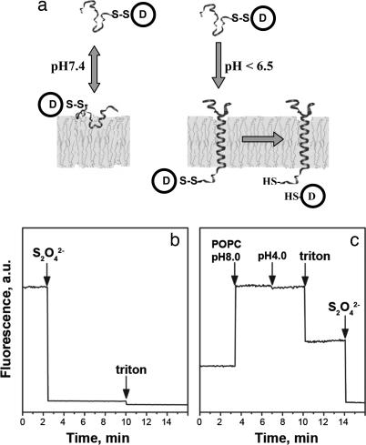
Fig. 1.
pHLIP insertion and topology. (a) Schematic diagram of cargo molecule delivery into a cell. At physiological pH, the peptide–cargo conjugate interacts weakly with a membrane. At low pH, the peptide forms a transmembrane helix with its C terminus inserted in the cytoplasm. Reduction of the disulfide bond releases a drug. (b) The topology of the pHLIP peptide in a lipid bilayer was determined by using the NBD–dithionite quenching reaction. The fluorescence signal of NBD attached to the N terminus of peptide was monitored at 530 nm when excited at 470 nm. Sodium dithionite was added to the NBD peptide inserted into POPC large unilamellar vesicles. (c) POPC large unilamellar vesicles containing dithionite ion inside were added to the NBD peptide at pH 8.0, and then decreasing the pH triggered insertion of the peptide in liposomes. Triton X-100 was used for the disruption of liposomes. The concentration of the peptide used in the experiments was 7 μM.

