
Figure 6.
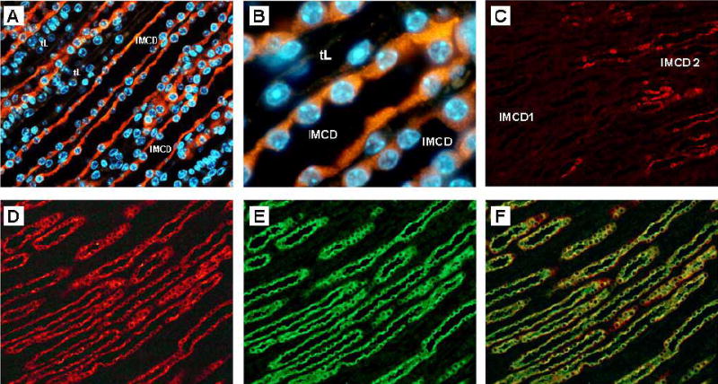
Localization of transgene in kidney inner medulla by immunohistochemistry. A; Low power magnification shows expression of β-Gal transgene (red) is confined to the cells of the IMCD and is not apparent in the thin descending limbs of Henle’s loop (tL). Nuclei are stained blue. B; High power magnification demonstrates that β-Gal staining (red) is apparent in the cytoplasm of the IMCD principal cells. C; Staining is localized to the IMCD2 and IMCD 3 regions, with little staining of the base of the inner medulla, IMCD1. Colocalization studies with an anti-aquaporin 2 antibody demonstrate that β-Gal staining (D, red) and aquaporin 2 staining (E, green) are colocalized in IMCD principal celIs, F.