
Figure 7.
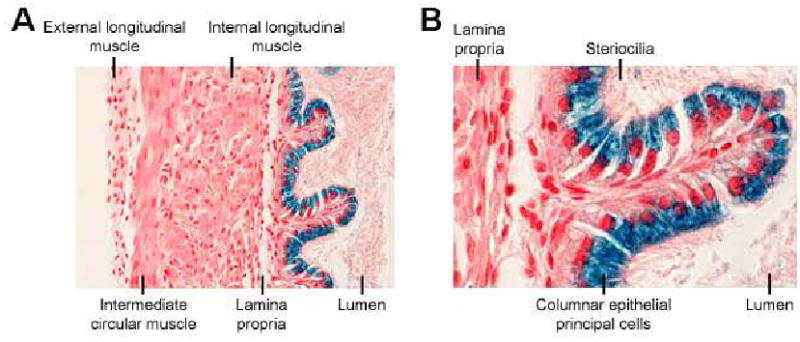
Localization of β-Gal enzyme activity in the vas deferens. X-Gal staining of the vas deferens demonstrated that β-Gal activity (blue staining) was observed in cells surrounding the tubule lumen (A). At higher magnification, staining was clearly localized to the columnar epithelial principal cells (B).