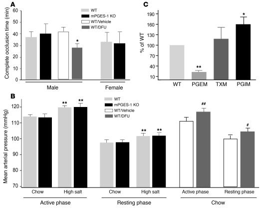
Figure 5. Impact of mPGES-1 disruption on thrombogenesis, blood pressure regulation, and eicosanoid biosynthesis.
(A) Deletion of mPGES-1 failed to alter the time to thrombotic carotid artery occlusion after photochemical injury, while it was accelerated by the PGHS-2 inhibitor DFU in mice on a DBA/11acJ background (*P < 0.05). (B) MAP exhibited diurnal variation in mPGES-1 KO and WT littermates on a mixed DBA/11acJ × C57BL/6 background. MAP was averaged over 4 days for 12 hours dark (active phase) and light (resting phase) periods. MAP was higher during active phase, and a high-salt diet elevated pressure similarly, a mean 6% in both groups (**P < 0.01). Oral DFU administration (10 mg/kg/d) for 21 days increased MAP in both the active (##P < 0.01) and resting (#P < 0.05) phases compared with vehicle-treated animals. (C) Urinary PGEM was lower (**P < 0.01) and PGIM was higher (*P < 0.05), while TXM was unaltered in male mPGES-1 KO mice compared with gender-matched WT littermates on a DBA/11acJ background.

