
Figure 3.
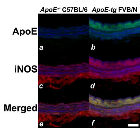
Immunodetection of apoE and iNOS after vascular injury in genetically-modified mice. Immnuofluorescent analysis of injured arteries from C57BL/6 apoE−/− mice (a,c,e) and apoE transgenic FVB/N mice (b,d,f) 24 hr after endothelial denudation. The sections were stained with fluorescent antibodies against apoE (a,b) or iNOS (c,d) to yield green and red signals, respectively. Panels e and f show the overlay of apoE (green) and iNOS (red) signals. Note that colocalization of signals appeared in yellow was detectable only in the medial smooth muscle cell layers of the arteries in FVB/N-apoE transgenic mice (f) but not in arteries of the C57BL/6 apoE-null mice (e). Scale bar represents 20 μm.