
Figure 4.
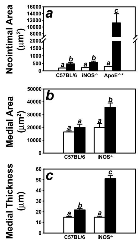
Morphometric quantification of control (open bars) and injured (filled bars) carotid arteries from C57BL/6 wild-type and iNOS-defective NOS2−/− mice. Panel A shows neointimal area of the injury carotid arteries of C57BL/6 wild type and iNOS-deficient mice in comparison with the neointima formed in the injured arteries of apoE-null (apoE(−/−) mice (* reported in ref 8). The Neointimal areas were determined by subtracting the luminal area from the area encircled by the internal elastic lamina. Panel B shows the morphometric data for medial area, which was calculated as the area encircled by external elastic lamina minus the area encircled by the internal elastic lamina. Panel C shows the mophometric data for medial thickness which was calculated as the average linear distance between the internal and external elastic lamina measured in four places at 90° apart. The data represent the mean ± SEM from ten C57BL/6 and ten iNOS(−/−) mice. Bars with different letters in each graph indicate significant difference at P < 0.05.