
Figure 6.
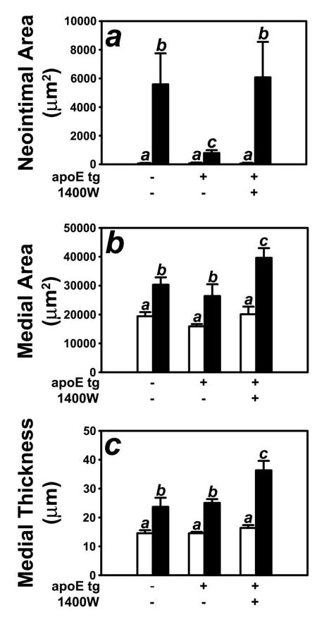
Morphometric quantification of control (open bars) and injured (filled bars) carotid arteries from FVB/N wild type and apoE transgenic mice given daily injections of saline or the iNOS inhibitor 1400W. Panel A shows neointimal area of the injury carotid arteries of apoE transgenic mice with or without 1400W. Neointimal area was determined by subtracting the luminal area from the area encircled by the internal elastic lamina. Panel B shows the morphometric data for medial area, which was calculated as the area encircled by external elastic lumina minus the area encircled by the internal elastic lamina. Panel C shows the morphometric data for medial thickness which was calculated as the average linear distance between the internal and external elastic lamina measured in four places at 90° apart. The data represent 10 mice from each group. Bars with different letters in each graph indicate significant difference at P < 0.05.