
FIG. 3.
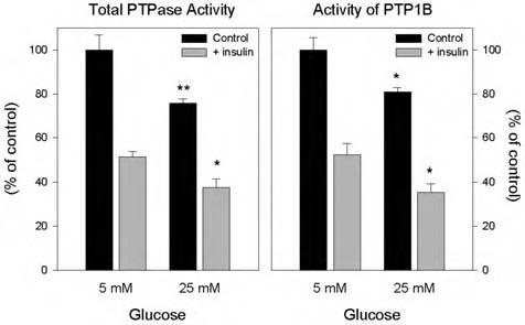
Effect of high glucose on the insulin-induced reduction of PTP activities in 3T3-L1 adipocytes. Total cellular PTP activity (left panel): Following overnight serum starvation, 3T3-L1 adipocytes cultured in 25 mM glucose for 8 days were treated with 100 nM insulin where indicated, then snap-frozen in liquid nitrogen, and lysed within the anaerobic chamber. To measure total PTPase activity, equal amounts of lysate protein (30 μg) were incubated in a final volume of 100 μl at 37°C for 30 min in reaction buffer containing 10 mM pNPP (Sigma) and 2 mM EDTA in 20 mM MES at pH 6.0 without dithiothreitol. The reaction was stopped by the addition of 900 μl of 0.2 M NaOH, and the absorption was determined at 410 nm. Activity of immunoprecipitated PTP1B (right panel): Using equal amounts of lysate protein (500 μg), under anaerobic conditions, lysates were precleared with nonimmune IgG and trisacryl protein G, followed by isolation of PTP1B by immunoprecipitation with a monoclonal antibody directed at a C-terminal epitope that preserves its enzymatic activity (Ab- 2). PTP1B activity was measured in washed immunoprecipitates as indicated above by hydrolysis of pNPP. *p < 0.5, **p < 0.001, compared with the corresponding samples in 5 mM glucose.