
FIG. 6.
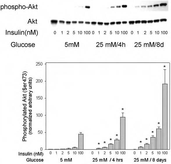
Effect of high glucose on insulin-stimulated Akt Ser473 phosphorylation in 3T3-L1 adipocytes. Serum-starved 3T3-L1 cells were incubated in the indicated glucose condition, stimulated for 5 min with the indicated concentrations of insulin, and samples were processed as described in the legend to Fig. 4. The western blots were probed with phospho-Akt (Ser473) antibody, followed by stripping and rehybridization with antibody to Akt protein for normalization. The asterisks indicate significant differences from the control cells cultured in 5 mM glucose (p < 0.05), as described in the text.