
FIG. 7.
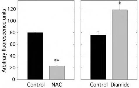
Effect of NAC or diamide on H2O2 production in 3T3-L1 adipocytes under high glucose conditions. NAC (left panel): Fully differentiated 3T3-L1 adipocytes maintained in 25 mM glucose medium were treated with 10 mM NAC for 1 h prior to measurement of H2O2 production by loading with DCF-DA (10 μg/ml) for 10 min followed by confocal fluorescence microscopy. Confocal images were quantitated using Scion Image software. **p < 0.001 versus control. Diamide (right panel): This experiment was performed as described for NAC, except that the cells were treated with diamide (30 μM) for 1 h prior to measurement of cellular H2O2 production on confocal microscopy. * < 0.02 versus control.