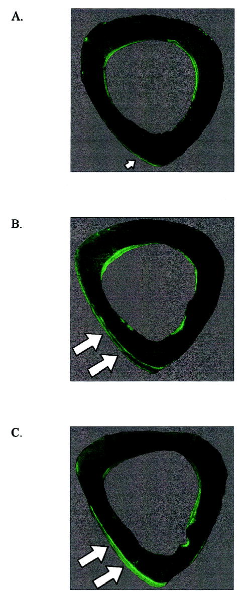
FIG. 3.

Composite fluorescent micrographs of the mouse midshaft illustrate (A) minimal periosteal bone formation in response to the low-magnitude regimen, and (B) substantial periosteal bone formation stimulated by the high-magnitude and (C) rest-inserted regimens (arrows). Endocortical bone formation was similar for each group and no different from that observed in the intact contralateral tibia.
