
Figure 1.
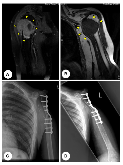
(A) T2-weighted and (B) T1-weighted coronal magnetic resonance images (MRI) of a 43 year old man presenting with osteosarcoma involving the proximal humerus (arrows). (C) Antero-posterior and (D) lateral post-operative plain radiographs demonstrating the surgical management, which comprised of a proximal humeral resection and a claviculo-prohumeral reconstruction of the shoulder.