
Figure 4.
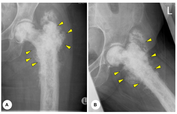
(A) and (B) Anteroposterior and lateral plain radiographs displaying typical pagetoid changes to the proximal femur with evidence of tumour extending beyond the cortex into the soft tissue (arrows). This 85 year old lady was managed conservative with radiotherapy alone.