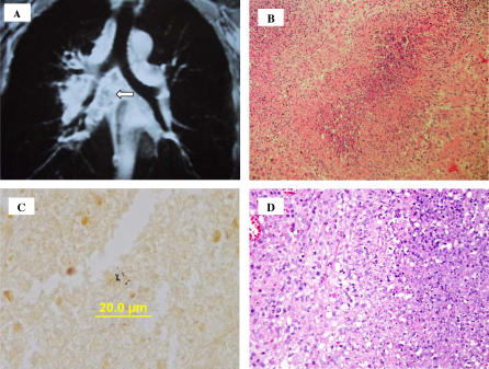
Figure 1. Clinical and Pathological Images of the Patient's Lymph Nodes.
(A) MRI of the chest showing adenopathy in the precarinal, subcarinal, and hilar regions. Central areas of diminished enhancement suggest necrosis (arrow).
(B) Removed cervical lymph node showing necrotizing granulomatous lymphadenitis with abscess formation (pyogranuloma).
(C) Warthin-Starry stain of the cervical lymph node (magnification 600×) showing coccobacillary organisms.
(D) Higher magnification H&E of a necrotizing granuloma. There is an area of neutrophils and cellular debris on the right, bounded by a poorly defined layer of palisaded epithelioid histiocytes. Outside this layer is a mix of lymphocytes and histiocytes.

