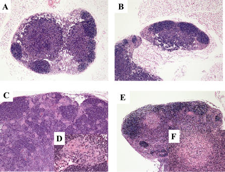
Figure 6. Histopathology of Mouse Lymph Nodes.
(A) Normal lymph node architecture of a gp91phox−/− uninjected control mouse (magnification 5×).
(B) Normal lymph node architecture of a gp91phox −/− mouse after 107 i.p. inoculation of Gluconobacter oxydans (magnification 5×).
(C) Moderate lymphoid hyperplasia and presence of atypical macrophages in the lymph node of a gp91phox −/− mouse after 106 intraperitoneal inoculation of the novel organism (magnification 5×).
(D) View of the epithelioid macrophages in the lymph node at 40×.
(E) Lymphadenitis in the cervical lymph node of a gp91phox −/− mouse after 107 i.p. inoculation of the novel organism (magnification 5×).
(F) View of the pyogranulomatous reaction in the lymph node at 20×.
(A,B,E,F) are from mice that were sacrificed at nine days post-inoculation. (C and D) are from a mouse that was sacrificed at 76 days post-inoculation.

