Figure 5.
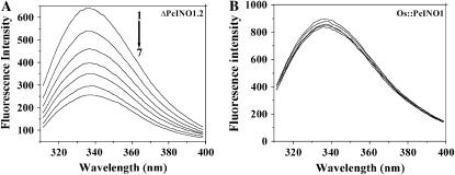
Fluorescence quenching of Trp. Trp fluorescence emission spectra of ΔPcINO1.2 (A) and Os∷PcINO1 (B). The excitation wavelength was 295 nm and emission was scanned between 300 to 400 nm. All proteins were in 20 mm Tris-HCl buffer, pH 7.5, containing 10 mm β-ME at a concentration of 0.15 mg/mL and the total reaction mixture was 1 mL. Line 1, Protein alone; line 2, protein + 100 mm NaCl; line 3, protein + 200 mm NaCl; line 4, protein + 300 mm NaCl; line 5, protein + 400 mm NaCl; line 6, protein + 500 mm NaCl; and line 7, protein + 600 mm NaCl. In all cases protein samples either in absence or presence of salt were incubated at 37°C for 10 min before subjected to emission scanning.

