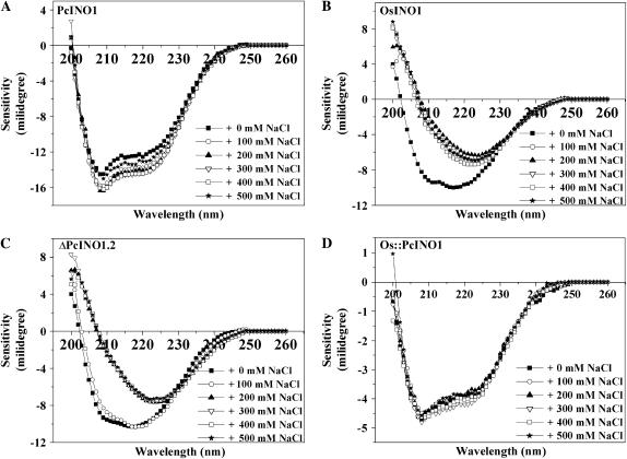
Figure 8.
Change in the secondary structure as revealed by CD spectra in OsINO1, PcINO1, and the mutant proteins in the presence of salt. CD profile of PcINO1 (A), OsINO1 (B), ΔPcINO1.2 (C), and Os∷PcINO1 (D) in presence and absence of increasing concentration of NaCl at room temperature. All the proteins were in 5 mm sodium phosphate buffer and approximately 0.1 mg/mL protein was used in each experiment. Spectrum was scanned between 260 to 200 nm using 2 mm pathlength cuvette in total 1.5 mL reaction volume. In all experiments, protein samples were incubated for 10 min at 37°C either in absence or presence of increasing concentration of added NaCl.

