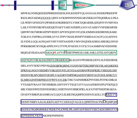
Figure 5.
Location of the CRE1 mutation in habituated calli. Schematic of the CRE1 protein domains was generated from the amino acid sequence at http://smart.embl-heidelberg.de/. Blue rectangles, Transmembrane domains. Green square/green box, HK domain. Green triangle/black underscoring, Catalytic domain. Blue pentagon/blue box, RR domain. The conserved His and Asp residues within the HK and RR domains, respectively, are depicted by a larger font size. The location of the amino acid change in habituated calli (F453L) is shown in red.

