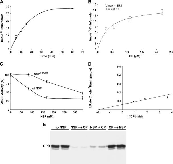
Figure 4.
NSP can inhibit AtNSI activity in vitro. A, Time course analysis of AtNSI activity. NSI (20 nm) was incubated with 3H-acetyl-CoA and 1 μm CaLCuV CP at 37°C for the times indicated to establish the linear range for the reaction. Using these conditions, a 10-min incubation period was used for further kinetic analyses. B, Michaelis-Menten kinetic analysis of NSI activity. Vmax and Km were calculated by incubating 20 nm NSI with 3H-acetyl-CoA and 1 μm CaLCuV CP for 10 min. C, NSP inhibition of AtNSI activity in vitro. NSI (20 nm) was preincubated for 5 min at 4°C with increasing amounts of CaLCuV NSP or NSPE150G, which is impaired in its ability to bind NSI, following which 3H-acetyl-CoA and 1 μm CaLCuV CP were added and the reactions were incubated for an additional 10 min at 37°C. D, Lineweaver-Burke plot of AtNSI activity. NSI (20 nm) was incubated with 3H-acetyl-CoA and increasing amounts of CP. Reactions were stopped after 10 min. E, NSP is not a deacetylase. An order-of-addition study was performed, in which 20 nm NSI was incubated with 4 μm CaLCuV CP and 3H-acetyl-CoA in the presence or absence of 80 nm CaLCuV NSP. Pairs of lanes show autoradiographs for 12% SDS-PAGE analyses of 3H-acetate-labeled CP products from independent duplicate reactions. No NSP, NSI was incubated with 3H-acetyl-CoA and CaLCuV CP for 15 min at 37°C. NSP was not added to the reactions. NSP → CP, NSI was preincubated with CaLCuV NSP for 5 min at 4°C, following which 3H-acetyl-CoA and CaLCuV CP were added and the reactions were incubated for an additional 15 min at 37°C. NSP + CP, CaLCuV NSP and CP were added simultaneously to NSI and 3H-acetyl-CoA. Reactions were incubated for 15 min at 37°C. CP → NSP,: NSI was incubated with CaLCuV CP and 3H-acetyl-CoA for 15 min at 37°C, following which CaLCuV NSP was added and the reactions incubated for an additional 10 min. Arrowhead marks position of CaLCuV CP (approximately 32 kD). The two faster migrating bands are breakdown products commonly seen in preparations of viral CP (see Pascal et al., 1994). 3H-acetate incorporation in A through C was determined by TCA precipitation and scintillation counting.

