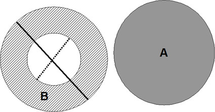
Figure 2.

Diagram of outer and inner diameters. Diagramatic draft of the measurement techniques applied to magnified cross-sectional images. The obvious round-shaped artery (A) and bronchus (B) pairs were identified and the outer (Do) and inner (Di) diameter of the bronchus were measured (solid and dotted lines respectively). WT% was calculated as [(Do-Di)/Do] × 100.
