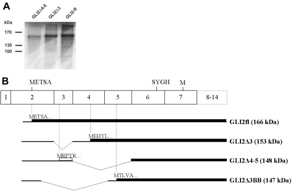
Figure 7.
In vitro translation of the GLI2 mRNA and its alternatively spliced forms. (A) SDS-gel analysis of [35S]-methionine labeled products translated with rabbit reticulocyte lysate. Constructs used are indicated on the top. Molecular weight marker positions are shown on the left. (B) Schematic representation of the translation products of the splice forms (shown on the right, with predicted molecular weight in parenthesis). mRNAs are shown by bold lines and their translation products are drawn by solid boxes. Open box represents translation in a different reading frame. The predicted N-terminal sequences (5 aa) are shown.

