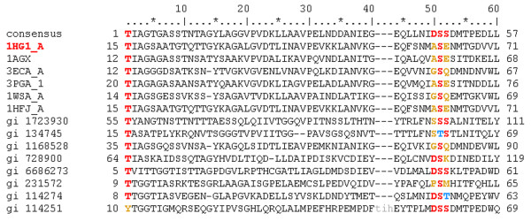
Figure 3.

A CDD domain family multiple alignment. All sequences from a CDD domain family are listed including the consensus. In addition, the sequence for the PDB protein from which the SMID interaction was derived is included, with its PDB code highlighted in red. Lowercase residues do not align with the consensus and represent insertions or deletions relative to the consensus. Small molecule binding site residues are mapped to the domain family sequences from the parent PDB sequence using the following colour-coding scheme: red for conserved residues, blue for similar residues and yellow for non-conserved residues. In cases where a binding site aligns to a gap in the consensus, conservation cannot be measured and thus no coloured residue is displayed. Note that some binding site residues may be highlighted in addition to those associated with the parent PDB sequence if there are redundant interactions from other PDB files with a similar binding site. This alignment has been truncated for clarity.
