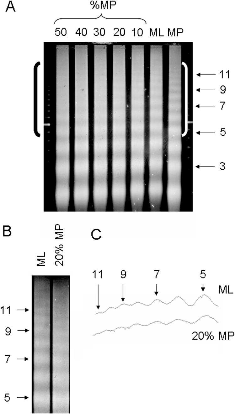
Figure 4.

Mixing experiment providing an estimation of the percentage of mouse liver chromatin that possesses a short repeat. (A) Nuclei from mouse liver (ML) or Matthiola petals (MP) were digested with MNase, and the purified DNA fragments were run on an agarose gel which was stained with ethidium bromide to visualize the nucleosome ladders from total genomic DNA for each sample (lanes ML and MP). The shorter nucleosome repeat (183 ± 5 bp) of the MP chromatin compared to the ML chromatin (195 ± 5 bp) is evident. The DNA from the two chromatin samples was mixed together in the proportions indicated (%MP) and analyzed on the same gel. Nucleosome oligomer bands for the MP chromatin are indicated. The brackets denote the upper region of the gel containing the oligomer DNA fragments greater than 5mers. (B) The 20% MP lane is shown adjacent to the ML lane, and the photograph was expanded for comparing the upper region of the gel. The lanes were precisely aligned using the 100 bp ladder markers immediately flanking the gel. (C) Densitometer scan of the ML and 20% MP lanes shown in (B).
