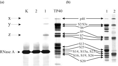
Figure 3.
Analysis of 40S ribosomal proteins cross-linked to the derivatives of HCV IRES by 1D PAGE in the presence of SDS. (a) Silver stained gel obtained in experiments with biotinylated IRES derivatives. Lanes 1, 2 and K correspond to the proteins isolated from the irradiated complexes of 40S subunits with RNA I, RNA II and RNA K, respectively. Lane TP40 corresponds to the electrophoregram of total 40S protein; positions of the 40S ribosomal proteins are indicated (35,36). (b) Analysis of proteins cross-linked to labelled RNA I in the binary complex with the 40S subunit. Lane 1, Coomassie stained gel; lane 2, autoradiogram of the gel.

