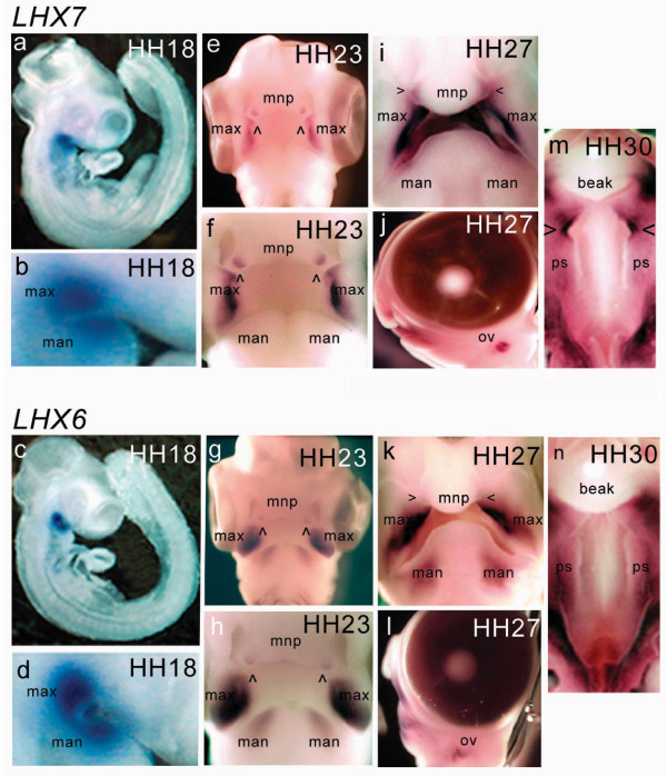
Figure 2.
Expression pattern of cLHX7 and cLHX6 in the developing chick embryo. cLHX7 (top panel) and cLHX6 (bottom panel) were restricted to the ventral extremities of the maxillary primordia and the rostral tip of the mandibular primordia before and after fusion of the maxillary primordia and medial nasal process during formation of the primary palate (a – i, k). From around HH27, cLHX6 expression was dispersed throughout the mandibular primordia (k). cLHX7 and cLHX6 expression was detected in the pre-fusion zone of the medial nasal process, prior to fusion with the maxillary primordia (e, f, g, h). The expression in the medial nasal process remained in the mesenchymal bridge of the beak after fusion (i, k). cLHX7 and cLHX6 expression was detected in the mesenchyme throughout the palatal shelves at HH30 (m, n). cLHX7 specifically displayed increased expression on the anterior tips of the developing shelves (m). Both cLHX7 and cLHX6 expression was detected in the otic vesicle from HH25 to HH30 (j, l). Abbreviations: max: maxillary primordia; man: mandibular primordia; mnp: medial nasal process; ov: otic vesicle; ps: palatal shelves.

