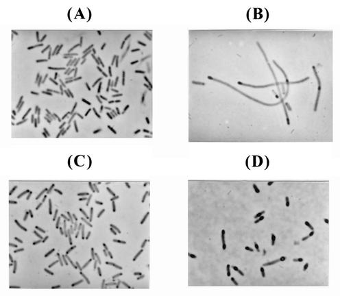
FIG. 4.
Morphologies of recombinant E. coli cells as observed by phase-contrast microscopy. (A and B) E. coli W3110 harboring pYKM-I1; (C and D) E. coli W3110 harboring pYKM-I1 and pACfAZ2. Panels A and C depict cells observed at 1 h after induction, while panels B and D depict those observed at 8 h after induction. Black inclusions inside the cells are inclusion bodies of IGF-I fusion protein.

