
Figure 2.
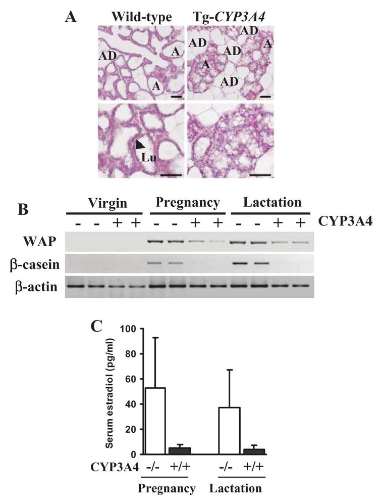
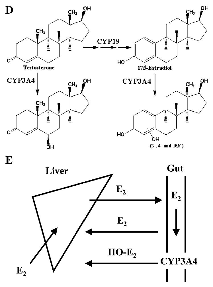
Impaired lactogenesis of CYP3A4-humanized mice associated with low estradiol levels. (A) Histological examination of mammary glands. In transgenic nursing mothers, it was sparsely filled with underdeveloped alveoli. In wild-type mice, the alveoli were fully distended by the accumulation of milk and minimal volume of adipose tissue (AD) was present. At higher magnification, the lumen (Lu) and epithelial cells (black arrow) of the alveoli are indicated. Scale bar: 50 μm. (B) Expression of milk protein genes in mammary glands as examined by RT-PCR. In both wild-type and CYP3A4-humanized virgin mouse mammary glands, whey acid protein (WAP) and β-casein were not detectable. During pregnancy and lactation, WAP and β-casein were abundant in wild-type mice and reduced or undetectable in transgenic mice. (C) Serum estradiol levels were significantly (*P < 0.05, n = 5 in each group) decreased in pregnant and lactating humanized mice. (D) Catabolism of testosterone and estradiol by CYP3A4. (E) Estradiol under enterohepatic circulation hydroxylated by intestinal CYP3A4.