Figure 6.

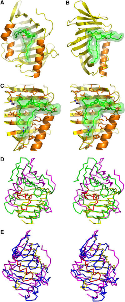
Hydrophobic cavity of LppX and structural comparison. A DIM molecule (green carbon atoms with carboxylate in red) is modeled into the cavity and is shown under a transparent surface, with LppX oriented as in Figure 5A (A) and rotated by 90° (B). (C) Close-up stereo view of the DIM molecule, oriented as in (B), showing the key aliphatic and polar residues (orange and with carbon atoms, respectively) that line the cavity. For clarity, the α-helices, α2 and α3, have been omitted in panels B and C. Overlay of LppX (yellow), oriented as in (A), and the crystal structure of (D) LolA (green, 1IWL) and (E) LolB (blue, 1IWN). Structural elements that significantly deviate in LppX are shown in magenta; those of LolA and LolB that differ from LppX cavity are in red.
